
mammograms
now browsing by tag
Mammograms VS. Thermograms For Breast Health
Mammograms vs. Thermograms for breast health. Have you ever had a mammogram? Have you ever had a thermogram? If you are over the age of 40 and female I am sure you have had a mammogram. A mammogram is the GOLD STANDARD for the mainstream, medical community for detecting breast 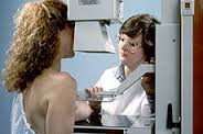 cancer. It entails a visit to the radiation department of your doctor’s office or hospital and you are led into a room where your breasts, one at a time, is squeezed between two metal plates and an x-ray is taken. The image is then read by a technician in another room. If anything suspicious is seen then more x-rays are taken and you are told that your doctor will call you with the results. Sometimes your doctor will request that you also have an ultrasound. If your tests are still unclear the next step is a needle biopsy or an operation to remove the lump called an lumpectomy. If it looks very concerning to the surgeon, the patient will be prepped before the operation that if the lump proves to be cancerous she can elect to have an mastectomy instead of just a lumpectomy. Can you image the STRESS this causes a women?!
(From http://www.thermographyonline.com/) Disclaimer: Thermography is utilized as an adjunctive imaging proce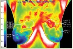 dure only and, as such, is not a replacement for or alternative to any other form of imaging. Since thermography only detects heat at the surface of the body, the technology cannot see into the cranial vault, thoracic or pelvic cavities, or deep into the body to visualize organs or bones. All thermography reports are meant to identify thermal emissions that suggest potential risk markers only and do not in any way suggest a diagnosis or treatment. Since a diagnosis cannot be made from an infrared image, thermal markers must be correlated by the patient’s treating physician with additional testing and procedures before a final diagnosis can be made.
Years ago I went in for my routine mammogram. I hated the entire experience. First, my anxiety level goes up when I just make the appointment because I fear what they will find. Then the actually experience of having your delicate breast tissue squished between two cold, metal plates can be painful and damaging in my opinion. If a suspicious spot is found then you will have to go through a number of addition x-rays causing more STRESS. The last time I had a suspicious area by a mammogram technician I had to position my body in several awkward positions for many different images of my right breast to be taken. All I was thinking was:
- This is uncomfortable.
- I am scared.
- How much additional HARMFUL x-rays and I getting pointed directly at my breast?
- Do I have CANCER?!
After that whole mess, I was sent home after I was told a doctor would review my images. Great! Now I get to go home and worry if I have cancer or not. Terrifying!
The next day my doctor called me saying that they needed MORE breast images so I had to go back in a for more x-rays. Are you kidding me? I did and no definitive answer was given so I was sent to have an ultrasound. At the ultrasound the suspicious area was measured and to my horror the technician practically snapped my head off when I asked her a simple question.
Back to my doctor’s office, she said that the suspicious area was determined to be a cyst, BUT she recommended that I see a surgeon and have the cyst aspirated so the cells could be examined under a microscope to see if it contained any precancerous cells. I sat in her office as she made the call to the surgeon’s office to schedule an appointment for me. I guess the receptionist on the other end of the phone asked if I was nervous because my doctor looked at me and asked me if I was nervous. By this time honestly I was no longer nervous because I had crossed over to PISSED. Are you serious? This is bull! The x-rays, the manipulation as my doctor tried to find the lump, the wait for the next appointment, the wait, wait, wait. I was beyond nervous and way into…this is just so irritating.
I did go to the surgeon’s appointment many days later, and the doctor did another ultrasound and determined that it was a cyst that would probably go away in a few months. If I wanted him to aspirate it (long needle into the breast) he would do so. I elected to come back in three months for another ultrasound. And in three months…the cyst was gone. Whew!
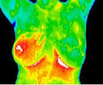
May I say, the entire process was HELL and I vowed to never go through that rigamarole again.That was my LAST mammogram to date. I decided to find an alternative way to monitor my breasts without the x-rays and without the squish of breast tissue. I found thermography. Thermography takes a image of your breasts and determines if there is anything suspicious by heat activity. A cancerous area will give off more heat than the rest of tissue and will show up on the image as a white spot. White being the hottest, red is warm showing inflammation, and blue is what you want to see. Go here for a better explanation. HERE
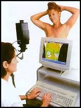 My appointment was in Sarasota, FL with Rita Rimmer, Certified Clinical Thermographer at Health Imaging. Rita was wonderful, friendly, and knowledgeable. She does the thermogram in a calm and quiet room with tranquil music playing softly in the background. I was told to take my shirt and bra off (Rita didn’t approve of my under-wire bra which restricts the lymph nodes.) and she aims the thermography camera/device at your breasts about 3 feet from where you sit and snaps via computer many pictures. While she is taking the pictures you can at the same time view the images on her computer screen, and she explains what is going on internally with your breast. Excellent! Much better than the ultrasound technician that snapped my head off.
(Source: From Rita Rimmer’s webpage) Thermography, although widely used in Europe and Australia, is beginning to make a comeback in the United States in the form of Digital Infrared Thermal Imaging (DITI).
DITI is more sensitive than previous thermographic technology and new digital thermal cameras are much smaller, more reliable, and significantly less expense than industrial cameras used in the 1970’s.
DITI is 100% safe, painless and requires no harmful radiation.
A Canadian Breast Cancer Screening Study demonstrated that thermography, when used in conjunction with mammography, can detect up to 95% of breast cancers.
I saw blue, yellow, green and red areas, 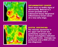 and the red was mostly around where the wire of my bra was just pressing against. Rita explained to me this is a sign of inflammation and explained what bras would be better for me to wear. She also explained that a thermogram will indicate a cancer/hot spot 10 years before it will show up on a mammogram. Rita gave me a lot of knowledge that day, and I am forever thankful to her for that. She relieved me from so much stress about whether I was going to find a cancerous lump the next time I did a breast self-examine or had a mammogram. In fact, I have never had another mammogram again and my family doctor is fine with that. Unfortunately, my health insurance does not cover the thermography and that is okay with me. I have no problem paying for it which at this time is over $100.00 but well worth it in my opinion. I think it reveals a lot more than a mammogram and knowledge is power. There are some health insurance plans that will cover it and I check from time to time to see if my personal plan has changed and will cover thermograms.
The traditional medical community has treated thermography as they did chiropractic care 20 years ago. They are not convinced it is reliable and they still rely on mammograms as the main diagnostic tool. For people who still want to have a mammogram I would recommend having both. I personally have friends who have had mammograms that have missed the cancer for years. (It is said that it takes 10 years for cancer to develop.) A thermogram can detect cancer 10 years before a mammogram.
Lets talk about what kind of radiation you receive from a mammogram.
General questions and comments on radiation risk
In large doses, radiation can cause serious tissue damage and increase a person’s risk of later developing cancer. The low doses of radiation used for imaging tests might increase a person’s cancer risk slightly, but it’s important to put this risk into perspective.
Point here, there is no minimum level of radiation that does not cause cancer. And to focus the radiation on your delicate breast tissue every year or two years…I will pass.
In conclusion, I determined that mammograms were not for me for the following reasons:
- Risk of cancer being caused by the radiation no matter how small the dose.
- Uncomfortable pain and pressure from the test.
- Unreliable results.
- False positives or positives false.
- The coldness of the entire process and the lack of communication.
- The waiting process for results.
- Having to go in for more tests and more x-rays for accurate diagnosis.
I am pro thermograms because:
- Same day results. (Your images are sent away for a professional review, too.)
- Personal interaction with the thermographer.
- You are given images for your personal records.
- No pain, no radiation.
- A clear explanation of your breast health and how to improve.
There are a couple of places to have a thermogram in the Sarasota area and if you Google thermography in your area you will most likely find one close to you. I recommend that you talk to your OBYGN or family doctor about whether to have a thermogram. Some are against it and some will recommend having both. As I said before, my doctors accept thermography and have no problem with me skipping the mammograms. I know some health practitioners reject thermograms and in some extreme cases insist that you have a mammogram every two years or they will not have you as a patient. Okay, everyone is entitled to their own opinion.
“Tell her Terry Ryan sent you! 941-330-9318”
If you decide to make an appointment with Rita Rimmer, here is her link at Health Imaging and please tell her Terry Ryan sent you. She is an awesome educator and I have tremendous respect for her, and thank her for making me feel more secure about my breast health.
Thank you for reading,
Terry Ryan, Health Blogger

Terry Ryan
Rita Rimmer, Clinical Thermographer Technician
Rita Rimmer’s FB page: https://www.facebook.com/rita.rimmer.3?fref=ts
Book on Thermography
 D5 Creation